
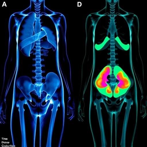
Body imaging techniques produce detailed pictures of the structures inside the body. These methods include MRI, CT, X-ray, and ultrasound, each capturing different types of internal detail. Radiologists rely on these tools daily, using them across nearly every area of care to observe the body. For people living with chronic conditions, imaging often becomes a regular part of ongoing management and follow-up. Here’s information on body imaging and how these techniques help support care plans for chronic conditions:
Visualizing the Body’s Internal Structures
Because each imaging method captures internal structures differently, each offers distinct benefits. X-rays pass radiation through the body, and denser tissues like bone absorb more of it, creating the contrast seen on the final image. CT scans build on this idea by combining many X-ray views into cross-sectional images, giving a layered view of organs, blood vessels, and soft tissues.
MRI uses strong magnetic fields and radio waves rather than radiation, highlighting soft tissues such as muscles, nerves, and the brain in fine detail. Ultrasound sends sound waves into the body and measures the echoes that bounce back, producing real-time images of organs and blood flow. Together, these methods give radiologists body imaging options suited to different:
- Body Tissues
- Diagnostic Questions
- Clinical Situations
Supporting Health Assessments
Chronic conditions often develop slowly; this is why tracking even small changes matters for effective medical care. Imaging supports this work by letting clinicians compare current findings with earlier scans. A radiologist can measure a structure’s size, note shifts in tissue appearance, or check whether a previously identified area looks different than before. This comparison helps care teams observe how a condition responds to a treatment plan.
For someone managing a long-term illness, repeat imaging can show whether findings remain stable and flag areas that need closer attention. Because different techniques reveal different details, care teams may combine methods to build a fuller picture of a patient’s status. Imaging also reduces the need for more invasive procedures in many cases. Instead of exploratory surgery, a scan can show internal structures clearly without incisions. This makes routine monitoring more practical for patients who require frequent assessments.
Diagnosing Health Conditions
When a person reports pain, swelling, or other changes, a radiologist can examine the affected area and describe what the images show. These findings give the broader care team specific information as they evaluate possible explanations to identify what is causing symptoms. The images produced during this process become part of a patient’s record. Different imaging techniques suit different diagnostic questions:
- Ultrasound: This technique is often recommended first because it is widely available and uses no radiation.
- CT Scans: These scans may be selected to quickly examine dense structures.
- MRIs: This imaging technique may be chosen when soft-tissue detail is the priority.
Care teams can compare later scans with earlier results to track how a situation evolves. This continuity supports the initial evaluation and the long-term follow-up that many chronic conditions require.
Learn More About Body Imaging
By pairing detailed images with clinical information, imaging helps care teams guide the next steps in treatment effectively. Body imaging gives radiologists and care teams a reliable way to observe internal structures, track changes, and support ongoing evaluations. Its range of techniques lets it address many tissue types and health questions. For people managing chronic conditions, imaging offers a consistent way to monitor their status over time. If you have questions about which imaging technique may fit your situation, contact a radiology practice for more information.